Cells, Free Full-Text
Por um escritor misterioso
Last updated 04 fevereiro 2025
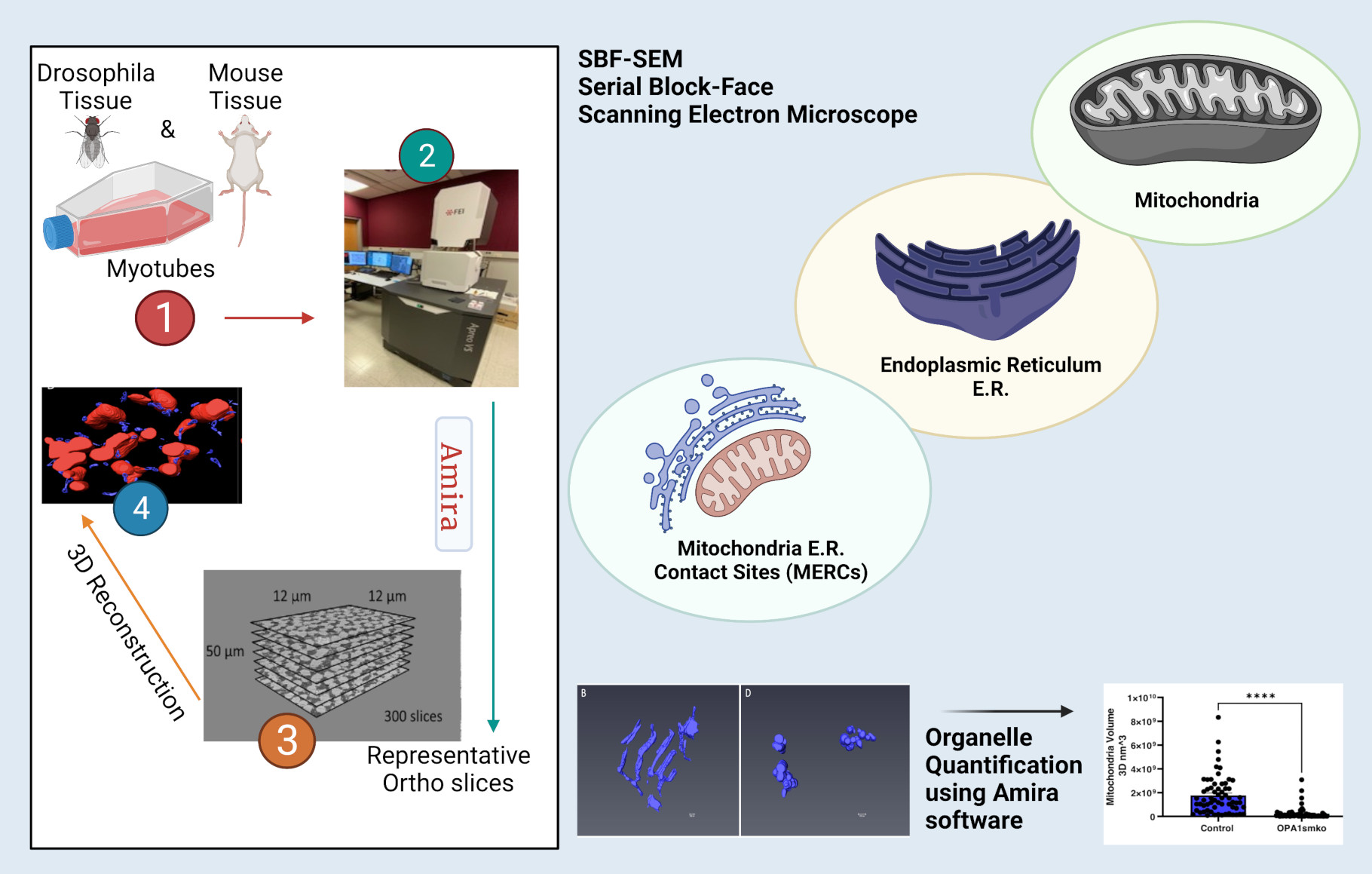
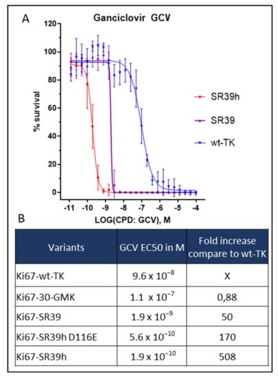
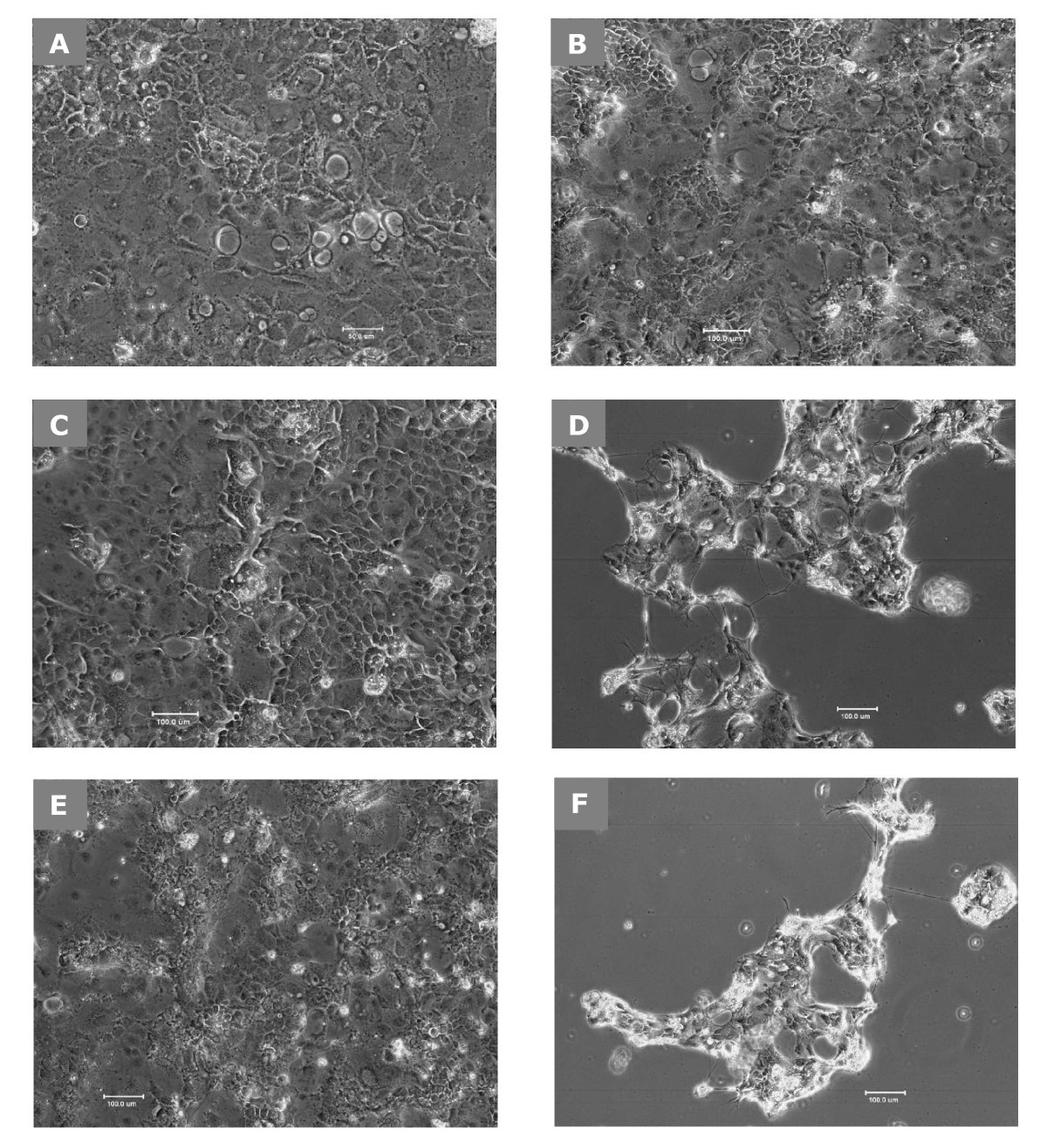
High-resolution 3D images of organelles are of paramount importance in cellular biology. Although light microscopy and transmission electron microscopy (TEM) have provided the standard for imaging cellular structures, they cannot provide 3D images. However, recent technological advances such as serial block-face scanning electron microscopy (SBF-SEM) and focused ion beam scanning electron microscopy (FIB-SEM) provide the tools to create 3D images for the ultrastructural analysis of organelles. Here, we describe a standardized protocol using the visualization software, Amira, to quantify organelle morphologies in 3D, thereby providing accurate and reproducible measurements of these cellular substructures. We demonstrate applications of SBF-SEM and Amira to quantify mitochondria and endoplasmic reticulum (ER) structures.

Cell-free Fetal DNA — A Trigger for Parturition
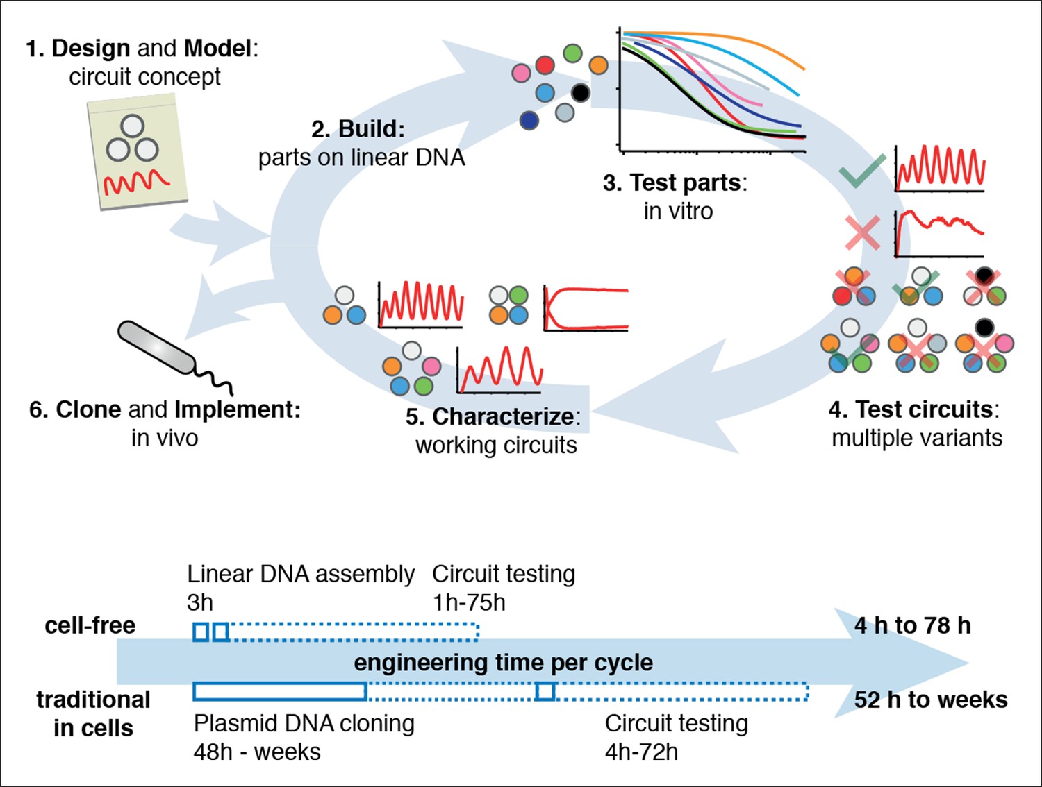
Rapid cell-free forward engineering of novel genetic ring oscillators

Sequencing of Circulating Cell-free DNA during Pregnancy
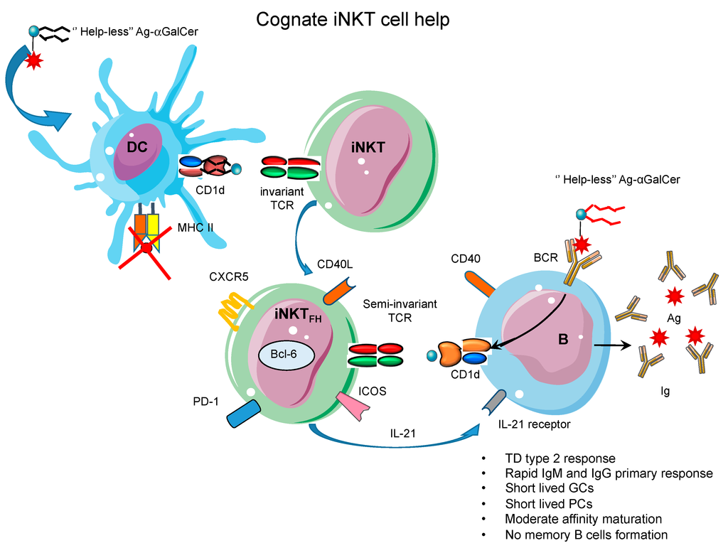
Antibodies, Free Full-Text

Cell-free synthetic biology: Engineering in an open world - ScienceDirect

MitoQuicLy: A high-throughput method for quantifying cell-free DNA from human plasma, serum, and saliva - ScienceDirect
Cells, Free Full-Text
Serial Number Alcohol 120 1.9 6 - Colaboratory

Cell-free expression and synthesis of viruses and bacteriophages: applications to medicine and nanotechnology - ScienceDirect

Cells, Free Full-Text
Lactobacillus delbrueckii ssp. bulgaricus B-30892 can inhibit cytotoxic effects and adhesion of pathogenic Clostridium difficile to Caco-2 cells, Gut Pathogens
Recomendado para você
-
 Mouse Accuracy - Mouse Accuracy and Pointer Click Training04 fevereiro 2025
 -
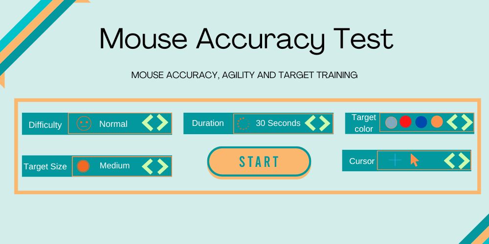 Mouse Accuracy Test Mouse Click Accuracy Precision04 fevereiro 2025
 -
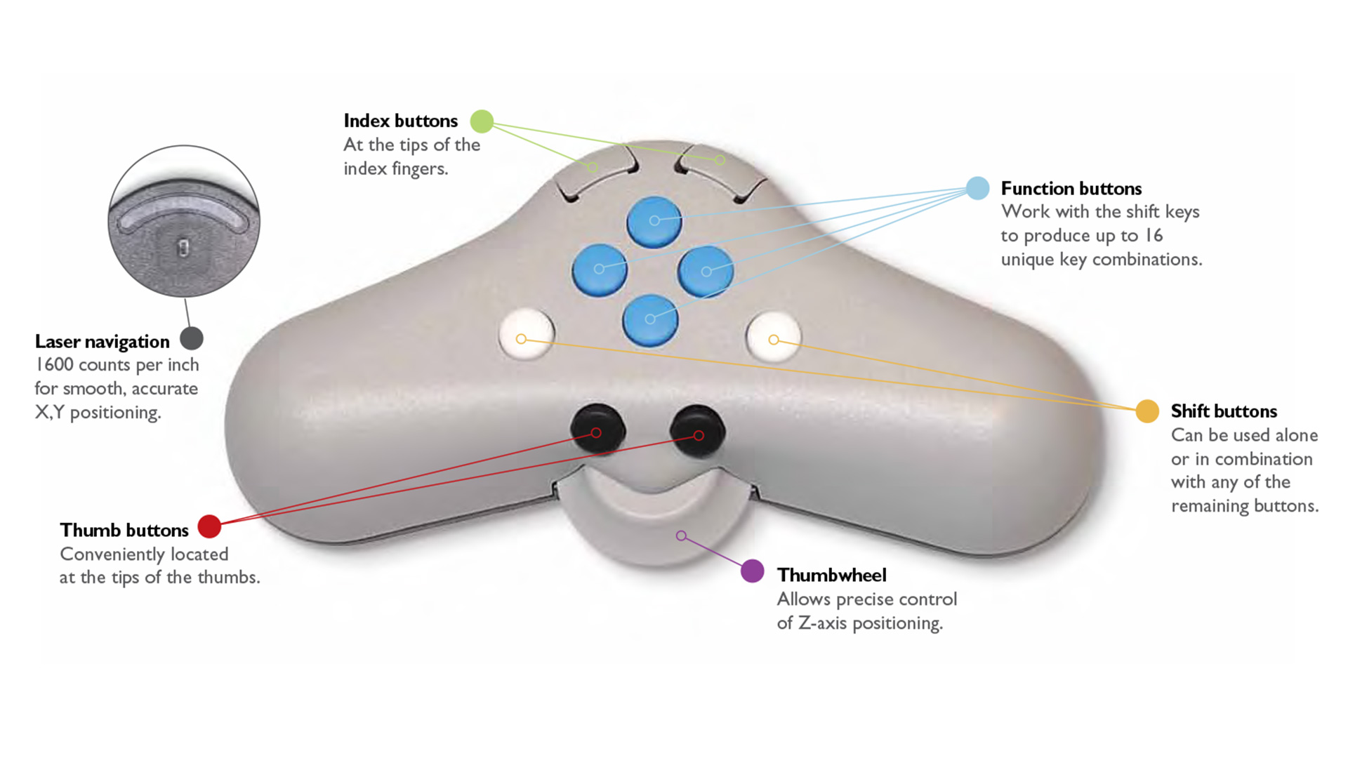 3D PluraView - 3D mouse, Professional graphics cards, photogrammetry gis04 fevereiro 2025
 -
 New 3-D printer is 10 times faster than commercial counterparts, MIT News04 fevereiro 2025
 -
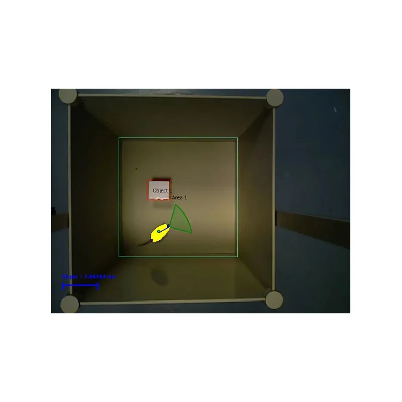 Novel Object Recognition Test 3D04 fevereiro 2025
 -
 Continuous Whole-Body 3D Kinematic Recordings across the Rodent Behavioral Repertoire - ScienceDirect04 fevereiro 2025
 -
 What is the Strongest 3D Printer Filament?04 fevereiro 2025
 -
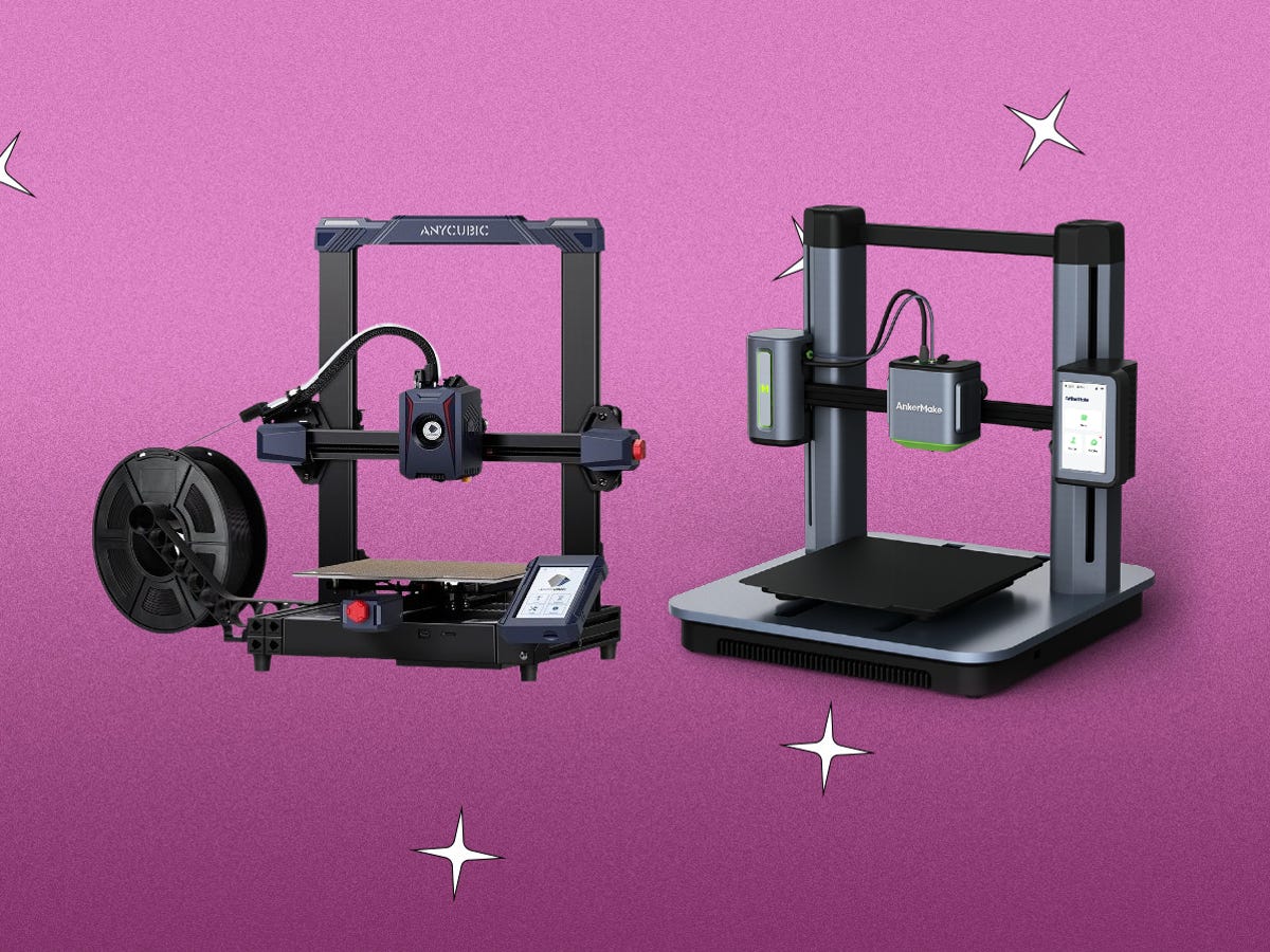 Best 3D Printer for 2023 - CNET04 fevereiro 2025
 -
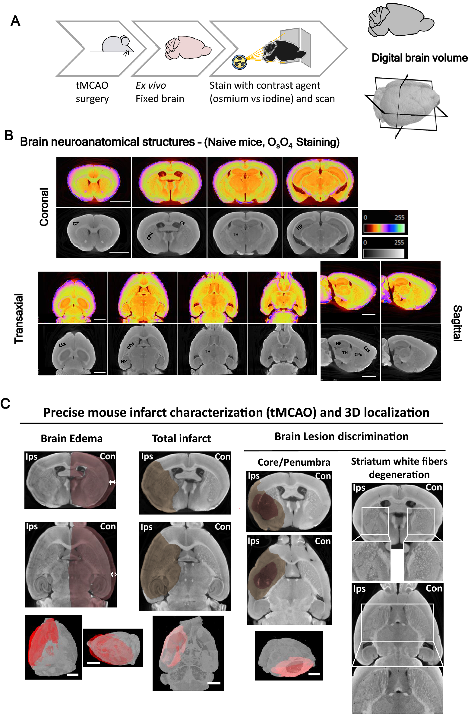 High-resolution micro-CT for 3D infarct characterization and segmentation in mice stroke models04 fevereiro 2025
 -
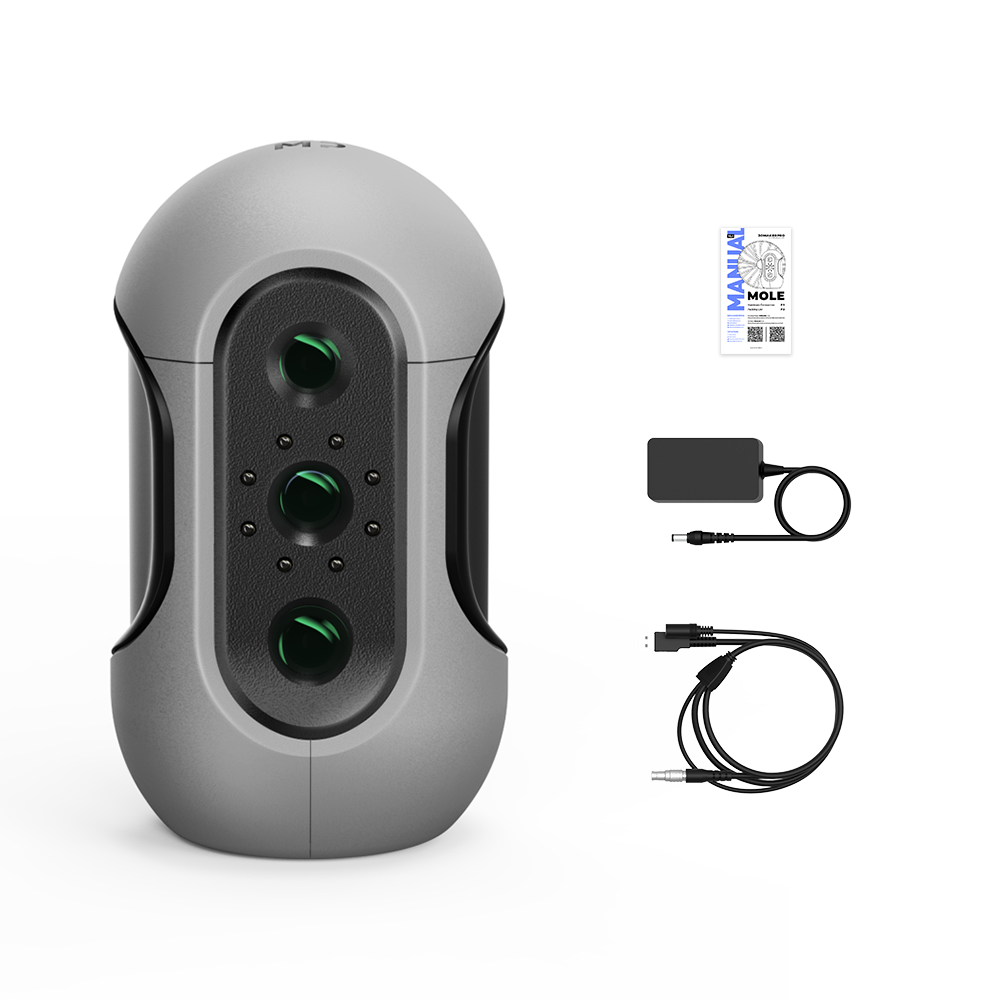 Mole 3D Scanner with Dedicated Mobile App04 fevereiro 2025
você pode gostar
-
 Menino triste sozinho no pôr do sol04 fevereiro 2025
 -
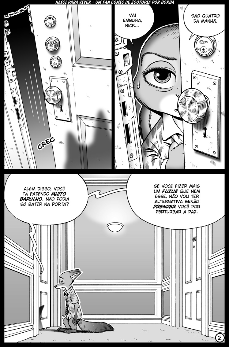 Nasci Para Viver - 02 by borba on DeviantArt04 fevereiro 2025
 -
 Season 6 Is FINALLY HERE!!!, Solaricks Overview & Review04 fevereiro 2025
 -
Bubble sort Algorithm Explained - Gadgetronicx04 fevereiro 2025
-
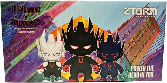 Limited Edition ZOTAC GAMING x Spider-Man: Across the Spider-Verse04 fevereiro 2025
 -
 The Ex-Navy SEAL Who Inspired 'Lone Survivor' on Learning to Recover - Men's Journal04 fevereiro 2025
 -
 SUPER Casa do Kame: setembro 201204 fevereiro 2025
 -
 Haverá uma nova evolução de Eevee? :: Poké Navegador04 fevereiro 2025
 -
 Ayanokouji Kiyotaka's Strategic Power Manipulation: Unveiling the 48 Laws — Eightify04 fevereiro 2025
 -
gratis.pixbet.com Concorrentes — Principais sites similares gratis.pixbet.com04 fevereiro 2025