Morphology of Leydig cells in the testes after in vivo MCP-1 treatment.
Por um escritor misterioso
Last updated 15 março 2025


Monocyte Chemoattractant Protein-1 (MCP-1): An Overview

Testicular macrophages are recruited during a narrow time window by fetal Sertoli cells to promote organ-specific developmental functions

Stem Leydig cells: Current research and future prospects of regenerative medicine of male reproductive health - ScienceDirect
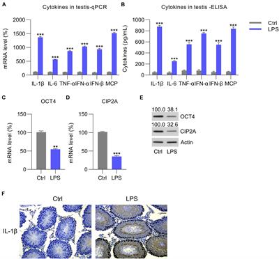
Frontiers OCT4 Represses Inflammation and Cell Injury During Orchitis by Regulating CIP2A Expression
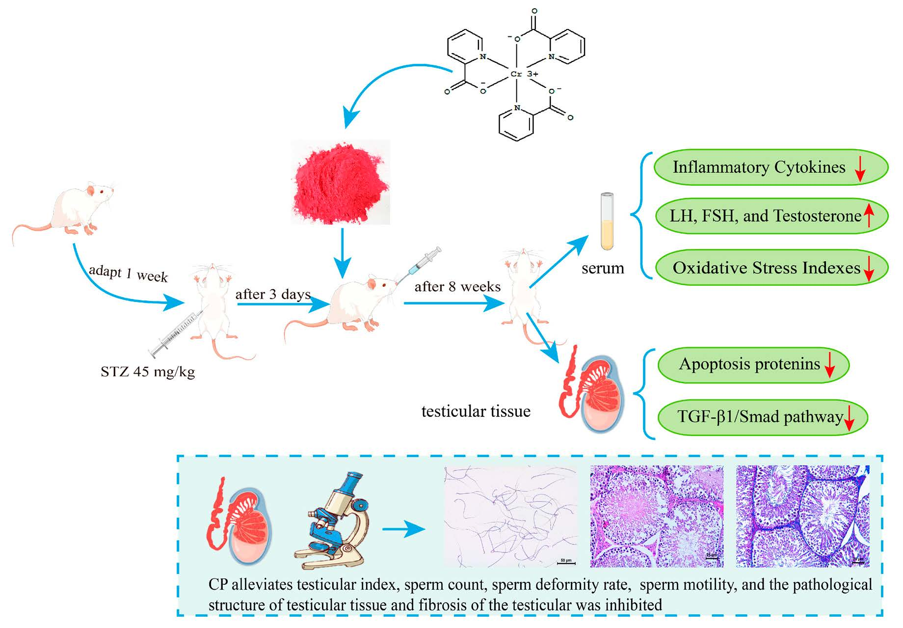
Molecules, Free Full-Text
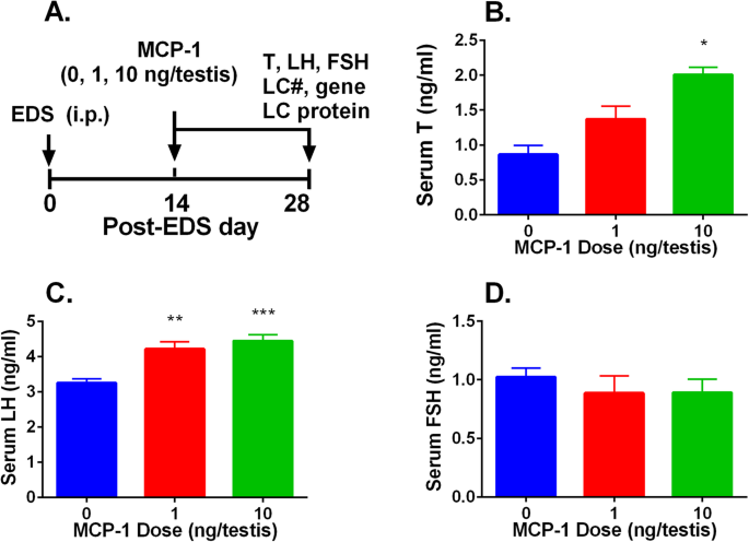
Monocyte Chemoattractant Protein-1 stimulates the differentiation of rat stem and progenitor Leydig cells during regeneration, BMC Developmental Biology

Fluoride-Induced Autophagy via the Regulation of Phosphorylation of Mammalian Targets of Rapamycin in Mice Leydig Cells

Testicular macrophages are recruited during a narrow time window by fetal Sertoli cells to promote organ-specific developmental functions

Morphology and cell number of Leydig cells after in vivo FGF1
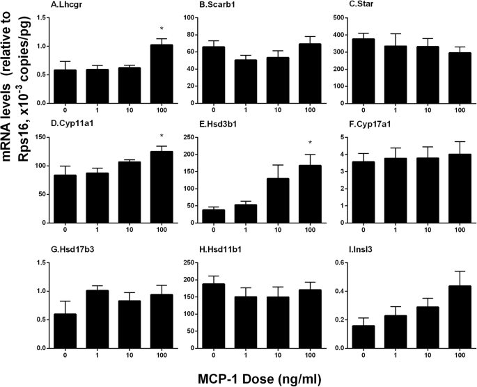
Monocyte Chemoattractant Protein-1 stimulates the differentiation of rat stem and progenitor Leydig cells during regeneration, BMC Developmental Biology
Recomendado para você
-
Faz o Teste - Ao Vivo - song and lyrics by Luan Estilizado, Wesley Safadão15 março 2025
-
 Blogueiro De Beleza Ao Vivo Maquiagem Diária Escovando O Teste Na Mão Usando O Telefone Celular No Tripé. Mulher Asiática Atraente, Influenciador Da Internet, Ou Vlogger Streaming Para Criar Conteúdo On-line. Foto15 março 2025
 -
 Teste Padrão Sem Emenda Do Vetor Vivo Do Córrego Para O Fundo Sem Emenda De Chromakey Da Substituição Video Do Blogue Para O Vlog Ilustração do Vetor - Ilustração de moderno, croma15 março 2025
 -
/i.s3.glbimg.com/v1/AUTH_08fbf48bc0524877943fe86e43087e7a/internal_photos/bs/2021/n/k/dg0dIJToA5v0aLmvm6vw/2015-11-19-batepapo41.jpg) Como testar transmissão ao vivo com um evento programado no15 março 2025
Como testar transmissão ao vivo com um evento programado no15 março 2025 -
/i.s3.glbimg.com/v1/AUTH_da025474c0c44edd99332dddb09cabe8/internal_photos/bs/2023/J/I/RRc9uPSAu4dWxgtdUqlg/tim.webp) TIM cria 'test-drive' para atrair clientes das rivais Claro e Vivo15 março 2025
TIM cria 'test-drive' para atrair clientes das rivais Claro e Vivo15 março 2025 -
/i.s3.glbimg.com/v1/AUTH_08fbf48bc0524877943fe86e43087e7a/internal_photos/bs/2022/I/R/Aow0BfRhKUd3kEimLWHw/print2.jpg) Como saber se a Vivo está fora do ar15 março 2025
Como saber se a Vivo está fora do ar15 março 2025 -
 Estamos Chegando a Imagem Escrita De Teste Ao Vivo Com Design Floral Ilustração Stock - Ilustração de arte, escrito: 21186803615 março 2025
 -
 Vai na Fé: Falsificando teste, Ben quase é pego por ligação: Dinheiro vivo15 março 2025
 -
 Mini Teste Ao Vivo 1550nm 20db Da Fibra Ótica Do Teste Ativo De15 março 2025
 -
Vivo X60 Pro 5G passa por teste de câmeras e bate Galaxy S21 Plus15 março 2025

você pode gostar
-
 Online Game Earn Money Website - Top, Best University in Jaipur, Rajasthan15 março 2025
 -
 I Love You, MrBeast15 março 2025
 -
 Mapa de Portugal: conheça suas regiões, distritos e concelhos - IE15 março 2025
 -
/cdn.vox-cdn.com/uploads/chorus_asset/file/24477359/Attack_on_Titan_Final_Season_THE_FINAL_CHAPTERS_Special_1_Still_1.jpg) Attack on Titan Final Season's last episode's release date has leaked - Polygon15 março 2025
Attack on Titan Final Season's last episode's release date has leaked - Polygon15 março 2025 -
 Is PDFCoffee.com Safe (2023)? - ANSWRED15 março 2025
 -
 Jogo De Cartas Pokémon Tcg Box De Batalha Pikachu - Copag15 março 2025
 -
Monster Halloween Doll Style D – Apps no Google Play15 março 2025
-
 Persona 5 Scramble The Phantom Strikers Limited Edition Korean15 março 2025
 -
 Random: Fan Makes Wii Menu Animations For Switch Games, And The Results Are Brilliant15 março 2025
 -
 Vector logo of the video game Minecraft. Steam (2702962)15 março 2025