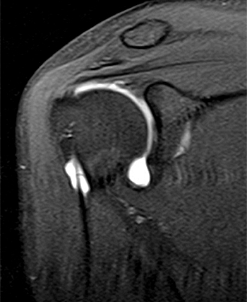
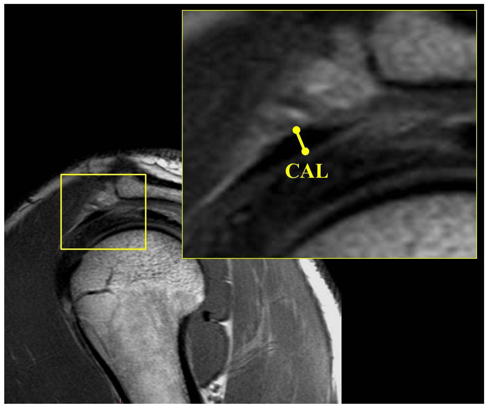
Typical magnetic resonance imaging scan showing the coracohumeral
Por um escritor misterioso
Last updated 21 setembro 2024


Magnetic resonance imaging of the shoulder
Subcoracoid Bursa

Alteration of coracoacromial ligament thickness at the acromial undersurface in patients with rotator cuff tears - ScienceDirect

The Primer for Sports Medicine Professionals on Imaging: The Shoulder - Nadja A. Farshad-Amacker, Sapna Jain Palrecha, Mazda Farshad, 2013
JCM, Free Full-Text

MRI of the Rotator Interval Capsule

Glenohumeral Instability
Radiological anatomy: X-ray, CT, MRI
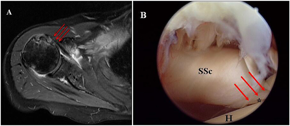
Frontiers Clinical efficacy and tendon integrity of patients with subscapularis tear by the technique of arthroscopic single external row repair

Pain related to rotator cuff abnormalities: MRI findings without clinical significance - Bencardino - 2010 - Journal of Magnetic Resonance Imaging - Wiley Online Library
Recomendado para você
-
 SCP 007- Abdominal Planet : r/SCP21 setembro 2024
 -
 SCP-007-J - Drawception21 setembro 2024
 -
 The SCP Foundation - Casual Cards - Yugioh Card Maker Forum21 setembro 2024
 -
 Confinement: SCPs / Characters - TV Tropes21 setembro 2024
 -
 SCP-007-RU-J - Суперзлодей21 setembro 2024
 -
 SCP基金会】SCP 007 J-松饼21 setembro 2024
 -
 SCP-S4S – SCP-007 Song Lyrics21 setembro 2024
 -
 LilyFlower's Workbench - SCP Foundation21 setembro 2024
 -
 SCP-S4S – SCP-484 Song Lyrics21 setembro 2024
 -
 049 and his lil brother 049-J how cute21 setembro 2024
você pode gostar
-
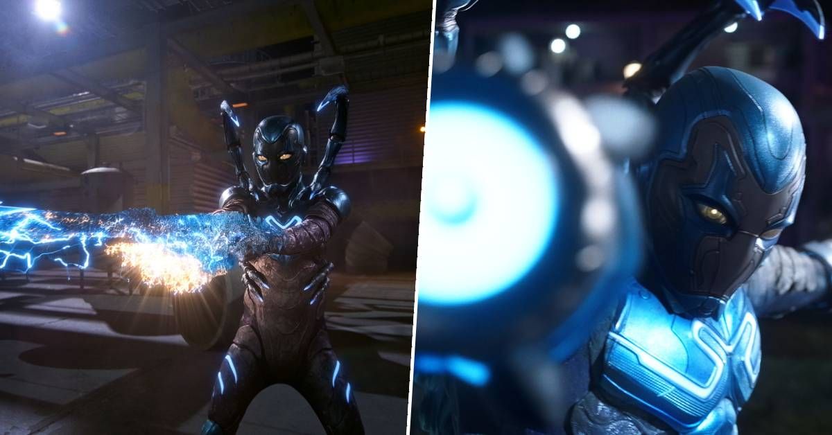 Is Blue Beetle in the DCU? Let's answer 2023's most confusing superhero movie question21 setembro 2024
 -
Chess players, what is the most satisfying checkmate? - Quora21 setembro 2024
-
 🤮 Face Vomiting Emoji, Vomit Emoji21 setembro 2024
 -
 Roupinha de Boneca - Vestido de Chapelzinho Vermelho21 setembro 2024
 -
 100-man no Inochi no Ue ni Ore wa Tatteiru ~Shide no Tabi-hen21 setembro 2024
 -
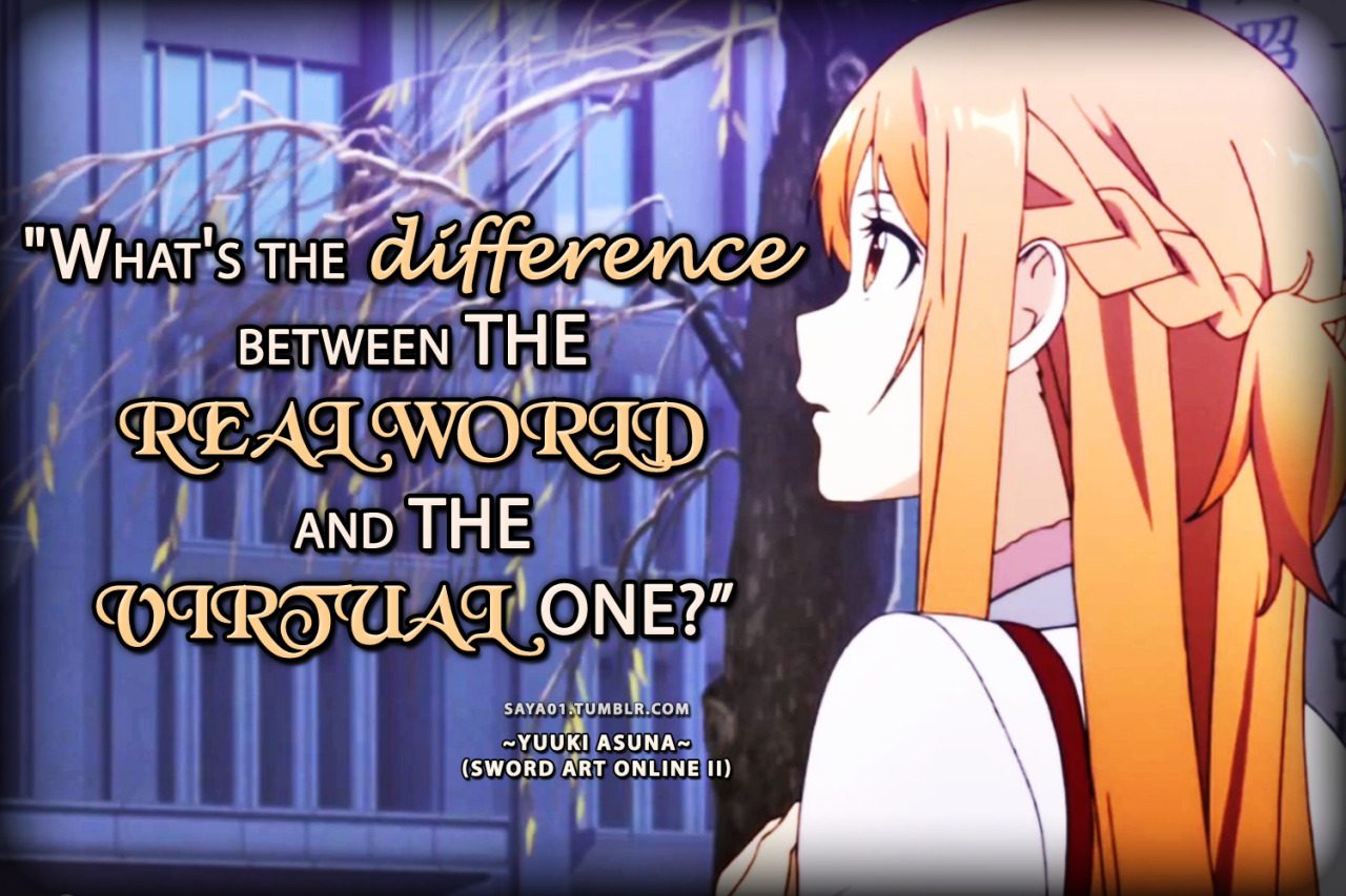 Anime & Manga Quotes — SWORD ART ONLINE II21 setembro 2024
 -
 Papel De Parede Adesivo Xadrez - Xadrez Vermelho Amarelo - Xadrez21 setembro 2024
 -
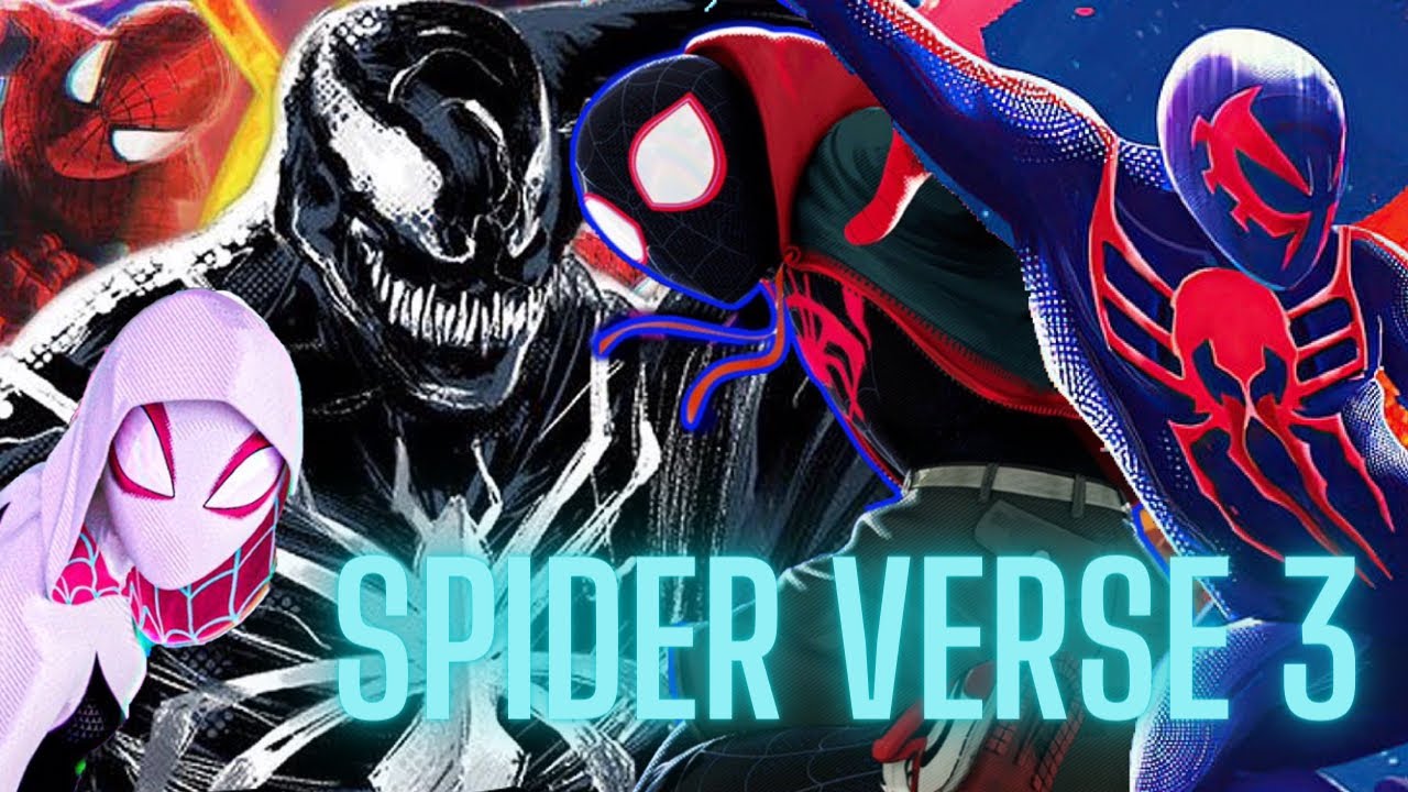 Spider Man Across the Spider Verse Part Two Release Date Confirmed21 setembro 2024
 -
 New Hunt: Showdown Roadmap for the Rest of 2023 and 2024 Detailed21 setembro 2024
 -
 Memes - Harry Potter - Filmes - Hary Potter - Página 6 - Criarmeme21 setembro 2024